RETINA

The retina is
the eye's film

Just as a damaged film makes photos blurry, retinal disease directly affects your vision.

WHY OLIM RETINA

Your sight depends on the retina—
catch problems early

Retinal detachment is an eye emergency. If a tear has formed but hasn't detached yet, laser can prevent detachment. When it's found changes the treatment.

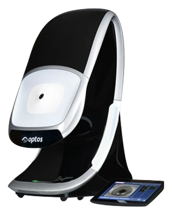
OPTOS DAYTONADiagnostic · Wide-field retinal imagingOPTOS DAYTONA
POINT 01

200-degree imaging
out to the periphery

OPTOS DAYTONA images 200 degrees of the retina without dilating drops, capturing the periphery—where retinal detachment often starts—in a single image.

  • OPTOS DAYTONA
  • No dilating drops
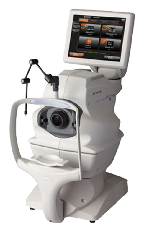
TOPCON 3D OCT-1Diagnostic · Retina & optic nerve OCTTOPCON 3D OCT-1
POINT 02

The macula,
layer by layer

TOPCON 3D OCT-1 optical coherence tomography shows cross-sections of the retina and optic nerve that can't be seen with the eye. We use it to diagnose macular degeneration and macular edema.

  • 3D OCT-1
  • Macular degeneration
  • Diabetic retinopathy
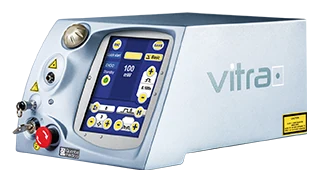
VITRA Monospot LaserSurgical · Retina & glaucoma laserVITRA Monospot Laser
POINT 03

At the tear stage,
laser can help

We perform retinal laser photocoagulation with the VITRA Monospot laser to prevent retinal detachment and manage new blood vessels in diabetic retinopathy and retinal vein occlusion.

  • VITRA Monospot
  • Laser photocoagulation
RETINA DISEASE

Five retinal conditions

Choose a condition to see its symptoms and treatment.

Retinal detachment

The retina lifts away from the wall of the eye. Your field of vision narrows and it feels like a dark curtain has fallen in front of your eyes.

Symptoms · Causes
  • Floaters, like flies drifting across your vision
  • Photopsia, flashes like a camera flash
  • Vision loss like a dark curtain coming down
  • Sudden loss of vision
Treatment
  1. 01Laser photocoagulation

    If there is a tear but no detachment yet, laser can help prevent detachment.

  2. 02Surgery

    If the retina has already detached, it is reattached with vitrectomy, scleral buckling, or gas or silicone oil injection.

AMSLER GRID

Macular degeneration self-test

If the lines look wavy, the center looks dark or empty, or the grid looks broken, get a detailed exam right away.

  1. Wear as usual

    Wear your glasses or reading glasses as usual and hold the grid at eye level in a bright place.

  2. Cover one eye

    Cover one eye and look at the center dot from about 30cm away.

  3. Look at the lines

    Notice how the lines look.

  4. Other eye

    Check the other eye the same way.

EQUIPMENT

Retinal exam equipment

VITRA Monospot Laser
Surgical · Retina & glaucoma laserVITRA Monospot Laser

A laser for retina and glaucoma treatment that provides non-invasive care through laser photocoagulation.

TOPCON 3D OCT-1
Diagnostic · Retina & optic nerve OCTTOPCON 3D OCT-1

Uses optical coherence tomography to image cross-sections of the retina and optic nerve that cannot be seen with the naked eye.

OPTOS DAYTONA
Diagnostic · Wide-field retinal imagingOPTOS DAYTONA

Captures 200° wide-field images out to the peripheral retina without dilating drops, for early detection of macular degeneration, diabetic retinopathy and retinal detachment.

WARNING SIGN

With these changes, see an eye doctor the same day

Retinal detachment is an eye emergency. Symptoms come on quickly, and delay can let the detachment spread.
If the changes below appear suddenly, don't wait for your next appointment.

TYPES

Retinal detachment falls into
three types by cause

Treatment depends on the type. If there's only a tear and no detachment yet, laser can prevent detachment; if it has already detached, surgery is needed to reattach the retina.

AMD TYPES

Macular degeneration: how do
dry and wet differ?

There are two main types of macular degeneration. Most cases are the slowly progressing dry type; in the wet type, abnormal blood vessels grow and vision can drop faster.

Dry macular degenerationWet macular degeneration
What happensThe macula thins and is damaged with age.Abnormal new blood vessels grow beneath the macula and damage it.
Rate of progressionOften progresses slowly over several years.Vision tends to decline faster than with the dry type.
Care · treatmentRegular exams track changes, and supplements (vitamins · minerals) are used depending on the stage.Anti-VEGF injections, photodynamic therapy (PDT) and other treatments slow vision loss.
WHO SHOULD CHECK

Who needs regular exams
even without symptoms

Diabetic retinopathy and early macular degeneration cause almost no symptoms at first. If any of the following apply to you, have a dilated retinal exam regularly even without symptoms.

Diabetes · blood vessels
  • If you have diabetes: a dilated exam at least once a year is generally recommended
  • If you have high blood pressure
  • The longer you've had diabetes, the higher the risk
Age · eye condition
  • Age 55 or older, or a family history of macular degeneration
  • You smoke
  • High myopia, or a history of eye surgery or major eye injury
  • You or a family member has had retinal detachment
EXAM

How a retinal exam
works

We put in drops to widen the pupils and examine the inside of the eye. If needed, imaging equipment captures the retina both wide and layer by layer.

  1. Vision · eye pressure

    We first check basic vision and eye pressure.

  2. Dilation

    We put in dilating drops and wait for the pupils to open.

  3. Fundus exam

    Through the dilated pupil, we examine the retina and optic nerve directly.

  4. Wide-field · OCT imaging

    Wide-field retinal imaging and OCT record the retinal periphery and cross-sections of the macula.

  5. Results explained

    We review the images with you and decide whether treatment is needed and when to follow up.

For a few hours after dilation, light may feel glaring and near objects may look blurry. Please don't drive yourself on exam day; bringing sunglasses helps.

DAILY CARE

Habits that protect the macula

Habits alone can't prevent retinal disease, but they may help lower the risk of macular degeneration or slow its progression.

RAISE YOUR VIEW. FOR LIFE.

One building.
Your eyes, for life.

From a child’s first Dream Lens to vision correction, eye-area care and presbyopia surgery. An eye clinic you’ll have a reason to return to at every age.

  1. Growing yearsChildren and parents

    We help manage myopia in growing eyes.

  2. 20s–30sVision correction

    A first step toward seeing without glasses or contacts.

  3. 30s–40sEye area and eye comfort

    Care for eyes and eye areas worn out by screens.

  4. 50s and upPresbyopia and eye health

    Presbyopia and cataract, plus the optic nerve and retina.

FAQ

FAQ

For anything else, ask us on WhatsApp.

I see things like little flies in my vision—are they all dangerous?

Floaters are common with aging, and if their shape or number doesn't change, they're usually not a big concern. But if they suddenly increase, or come with flashes of light or a shadow blocking your vision, it could be a retinal tear or detachment—get a detailed fundus exam right away.

I have diabetes, but my eyes feel fine. Do I still need an exam?

Yes. Diabetic retinopathy has almost no symptoms early on. With diabetes, a dilated exam at least once a year is generally recommended; discuss the exact interval at your visit.

If the Amsler grid looks normal, does that mean no macular degeneration?

The Amsler grid is only an indirect way to notice changes at home, not a diagnostic test. Early macular degeneration may have no symptoms, so regular eye exams are needed.

If one eye has macular degeneration, will the other eye get it too?

Advanced macular degeneration in one eye raises the risk in the other. Have both eyes checked regularly.

Can retinal detachment be prevented?

It's often caused by aging, so it's hard to prevent completely. But if it's found at the tear stage before detachment, laser can prevent detachment—so regular exams and prompt visits matter.

Can I drive right after a dilated exam?

For a few hours after dilation, light may feel glaring and vision may be blurry. Avoid driving on exam day and use public transport or have someone accompany you.


If your vision has suddenly changed

Retinal damage is hard to reverse once it's late. Get checked right away.