About 60
detailed tests
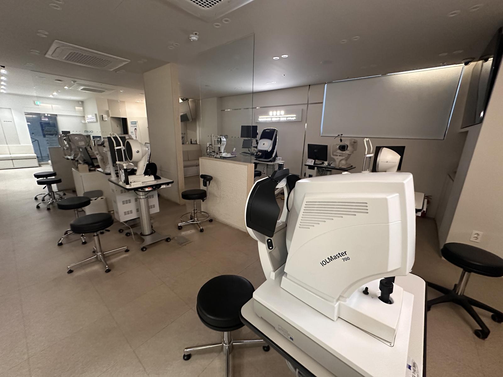
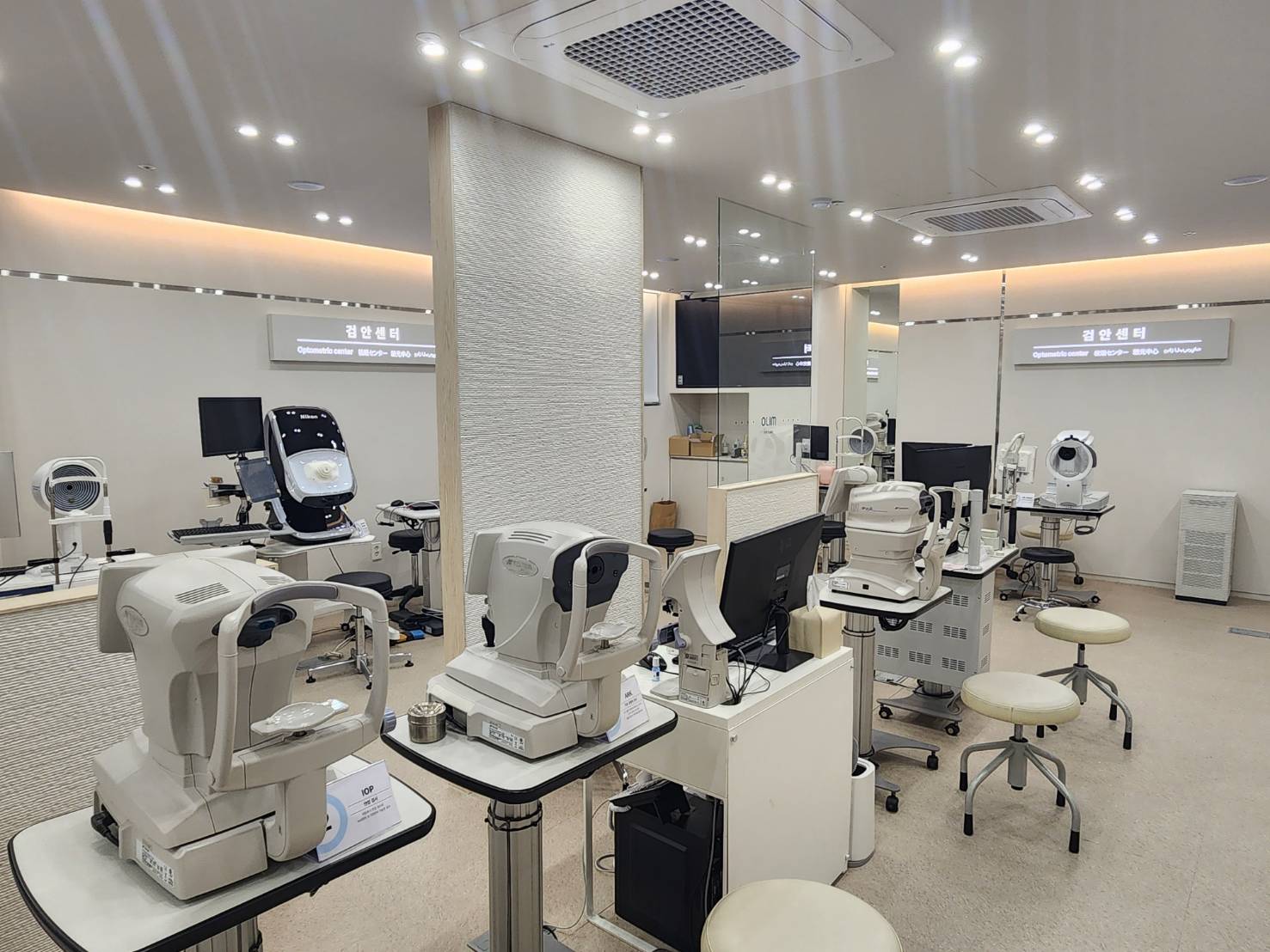
From vision and refraction to eye pressure, cornea, tear film, retina and optic nerve, we check with 12 diagnostic devices.
- About 60 tests
- 12 diagnostic devices
- Eye Exam Center

About 60 tests to check the whole eye.
Diseases that progress without pain, such as glaucoma and retinal disease, are often advanced by the time you notice them. The OLIM Eye Clinic Gangnam Eye Exam Center checks your whole eye with about 60 detailed tests.

From vision and refraction to eye pressure, cornea, tear film, retina and optic nerve, we check with 12 diagnostic devices.
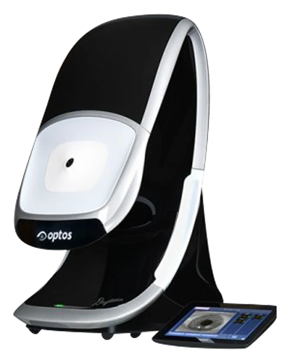
Diagnostic · Wide-field retinal imagingOPTOS DAYTONAOPTOS DAYTONA captures 200° wide images out to the peripheral retina without dilating drops, helping detect macular degeneration, diabetic retinopathy and retinal detachment early.
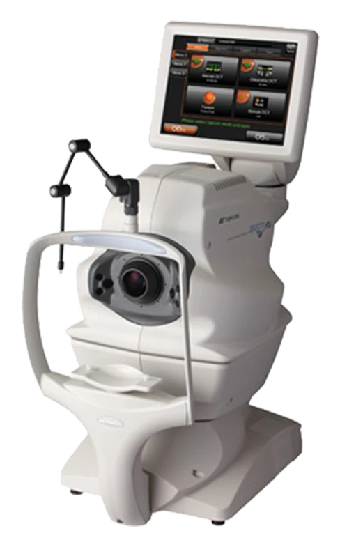
Diagnostic · Retina & optic nerve OCTTOPCON 3D OCT-1Refractive errors and lens side effects in your teens to 30s; presbyopia, cataract, glaucoma and diabetic retinopathy in your 40s; and macular degeneration too in your 60s. The conditions to check vary by age.
| Conditions checked | |
|---|---|
| Teens to 30s | Reduced vision such as myopia, hyperopia, amblyopia and astigmatism; eye injury, styes, conjunctivitis, hereditary glaucoma, cataract, dry eye, contact lens complications |
| 40s | Reduced vision such as presbyopia; dry eye, cataract, glaucoma, diabetic retinopathy, dry macular degeneration |
| 60s | Cataract, presbyopia, glaucoma, diabetic retinopathy, macular degeneration |

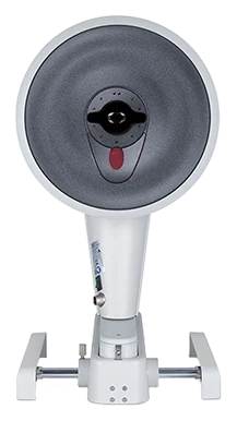
Analyzes corneal topography, thickness and shape to detect abnormal corneas such as keratoconus before surgery.

Uses optical coherence tomography to image cross-sections of the retina and optic nerve that cannot be seen with the naked eye.

Captures 200° wide-field images out to the peripheral retina without dilating drops, for early detection of macular degeneration, diabetic retinopathy and retinal detachment.

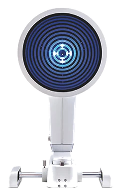
Images the meibomian glands and measures tear meniscus height, tear break-up time, lipid layer and redness.

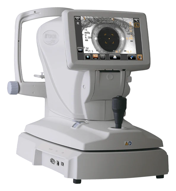
Measures eye pressure without touching the eye. Used for early glaucoma diagnosis and follow-up.

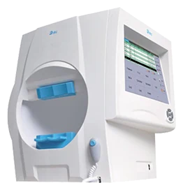
Measures the visual field by recording responses to small points of light shown in different positions.

A comprehensive eye exam is an overall check of your eye health by an ophthalmologist. Refraction, eye pressure, fundus, corneal thickness and dry eye tests are done according to your age and eye condition.
Even with good vision, there may be changes in the retina, optic nerve or visual field. That is why we look at several tests together.
| What it checks | Device used | |
|---|---|---|
| Vision · refraction | Degree of myopia, hyperopia and astigmatism, astigmatism axis, corneal curvature | TOPCON KR-800 |
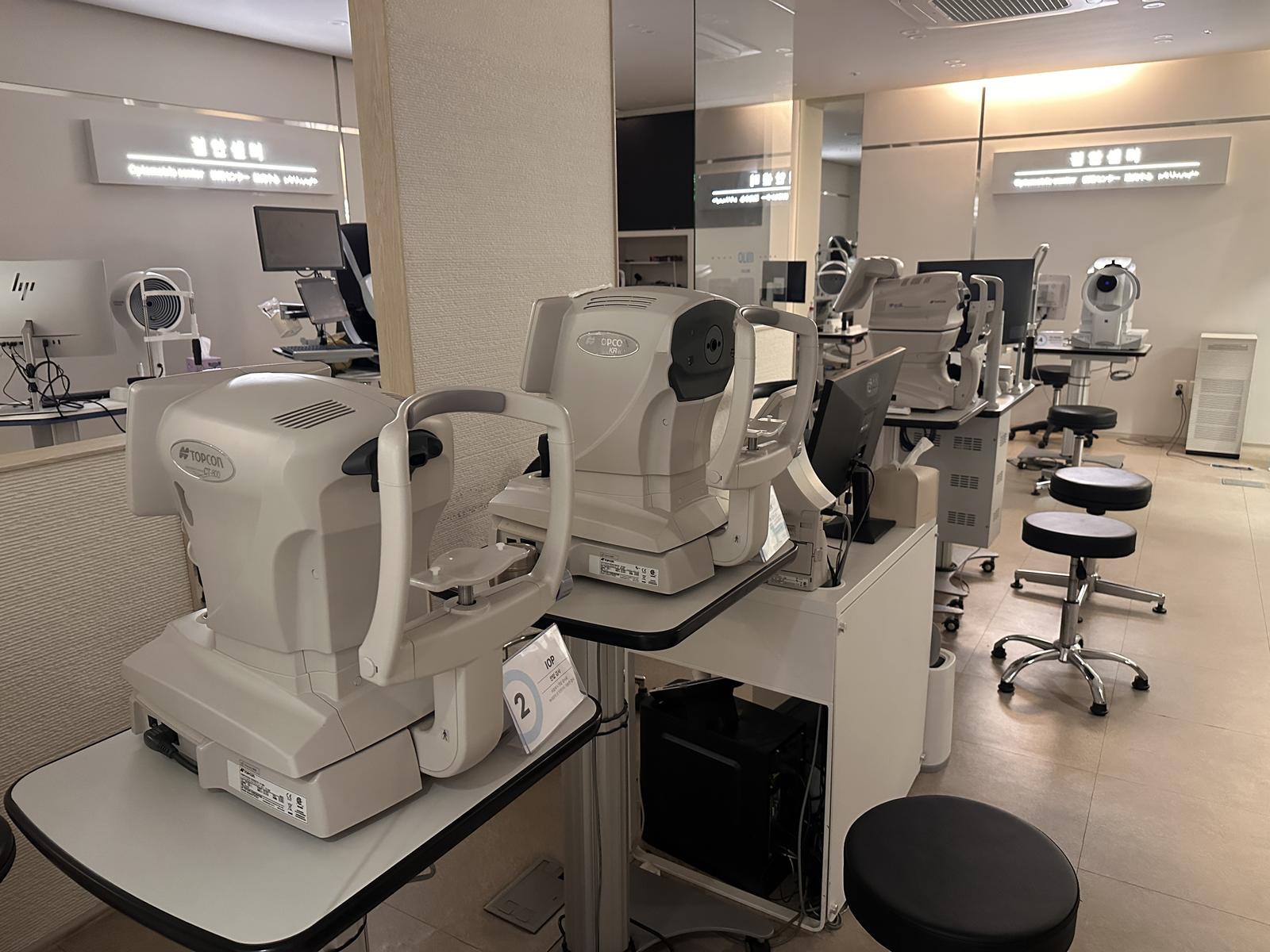
| Eye pressure | Pressure inside the eye. Used to detect glaucoma and track its course | TOPCON CT-800 (non-contact) |
| Visual field | Measures peripheral vision by your response to small lights in different positions | IRC IVS-201B |
| Wide-angle retinal imaging | Images out to the peripheral retina without dilating drops. Checks for macular degeneration, diabetic retinopathy and retinal detachment | OPTOS DAYTONA |
| Retina · optic nerve tomography | Cross-sections of the retina and optic nerve that can’t be seen by eye | TOPCON 3D OCT-1 |
| Corneal shape · thickness | Corneal topography and thickness. Checks for abnormal corneas such as keratoconus | Pentacam |
| Corneal endothelial cells | Condition of the cells on the inner cornea, and corneal swelling | TOPCON SP-1P |
| Tears · dryness | Tear meniscus height, tear film breakup time, meibomian glands, redness | OCULUS Keratograph 5M |
| Front of the eye | Direct microscope examination of the conjunctiva, cornea, lens and more | Slit-lamp microscope |
The order may vary depending on your eye condition and the tests you need.
We check your symptoms, the medicines you take, underlying conditions such as diabetes or high blood pressure, and eye diseases in your family.
Vision, refraction and eye pressure are measured.
The retina, optic nerve and cornea are imaged. If needed, drops that widen the pupil (dilating drops) are used.
The medical team explains your results and advises on any treatment needed and when to come back.
When using devices for long periods, rest your eyes by looking into the distance now and then.
Wear sunglasses outdoors to reduce UV exposure.
Diabetes and high blood pressure can also affect the retina and optic nerve.
Even without symptoms, have your eyes checked once a year.
From a child’s first Dream Lens to vision correction, eye-area care and presbyopia surgery. An eye clinic you’ll have a reason to return to at every age.
We help manage myopia in growing eyes.
A first step toward seeing without glasses or contacts.
Care for eyes and eye areas worn out by screens.
Presbyopia and cataract, plus the optic nerve and retina.
For anything else, ask us on WhatsApp.
Many eye diseases have no symptoms early on. In some, like glaucoma, your field of vision narrows before your sharpness does, so even if you feel you see well, a detailed exam is the way to confirm.
We recommend a detailed exam once a year. If you are 60 or older or have a family history of glaucoma, every 1–2 years; if you have diabetes or high blood pressure, generally at least once a year.
If drops that widen the pupil (dilating drops) were used, your vision may be blurry and glare-sensitive for a few hours, so please avoid driving yourself. You can ask before the exam whether dilation will be used.
Most involve sitting in front of a device and looking at a screen or light. Eye pressure is measured with a non-contact tonometer that sends a brief puff of air without touching your eye.
In your teens to 30s, we mainly check for reduced vision such as myopia and astigmatism, conjunctivitis, dry eye and lens side effects; in your 40s, presbyopia, cataract, glaucoma and diabetic retinopathy; and in your 60s, cataract, glaucoma, macular degeneration and more.

Book ahead and you can be examined without waiting.